Seeing the Unseen
Seeing the Unseen
Guangwei Hu, Nanyang Technological University, Singapore
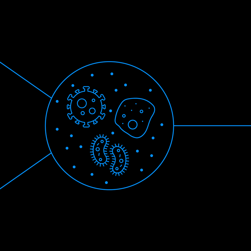
All around us, a hidden world exists that remains invisible to the naked eye. The adult human body has between 28 and 36 trillion cells spread over 400 cell types, all too small to be seen, except for egg cells. Microplastics litter the oceans like flecks of snow, remnants of the tens of millions of tons of plastic that enter Earth’s oceans every year. Some challenges become even more significant—or smaller depending on your perspective: nanoplastics, particles even smaller than microplastics, may result in even more critical health hazards. And fine particulate matter, much smaller than the width of a human hair, can pose a threat to both air quality and human health. Air pollution as a carcinogen is a topic of active study, with early research demonstrating a link between particulate matter and gastrointestinal cancers.
Visualizing such tiny, often transparent objects requires specialized tools that are bulky and confined to a laboratory setting. For example, differential interference contrast (DIC) microscopy can enhance the contrast in unstained, transparent samples in real time that are challenging to be observed with other methods. However, a traditional DIC microscope is a tabletop system that demands a large number of optical components for operation.
“The democratization of light microscopy is expected to provide individuals living in developing countries or remote areas with the capability to analyze microplastics in drinking water and air, as well as detect pathogens in biological samples.”
“The democratization of light microscopy is expected to provide individuals living in developing countries or remote areas with the capability to analyze microplastics in drinking water and air, as well as detect pathogens in biological samples.”
For the betterment of human health and the environment, researchers around the world are aiming to take advanced light microscopy techniques out of the lab and into the field. Some groups—including my own lab—are leveraging flat, ultrathin optical elements and consumer electronics such as smartphones to develop portable, miniaturized versions that rival their much larger counterparts. As devices become more portable and available to researchers—and even the public—we can expect to advance solutions more quickly.
The democratization of light microscopy is expected to provide individuals living in developing countries or remote areas with the capability to analyze microplastics in drinking water and air, as well as detect pathogens in biological samples. In places with limited medical and environmental monitoring facilities, improved accessibility of light-based technologies has the power to save lives.
Visualizing transparent samples
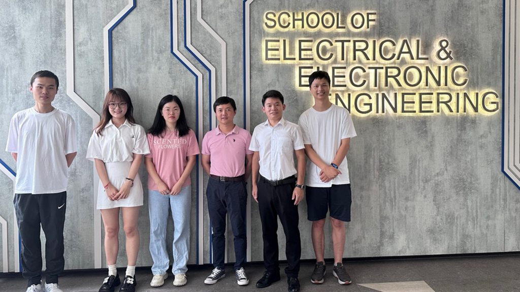
Hu (third from right) and his group at Nanyang Technological University, Singapore. Credit: Guangwei Hu
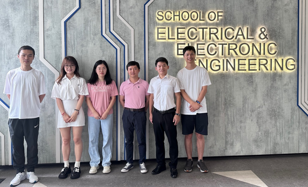
Hu (third from right) and his group at Nanyang Technological University, Singapore. Credit: Guangwei Hu
Because many cells in their natural state lack color and contrast, biologists who use conventional light microscopy based on absorption must introduce stains or fluorescent dye. However, the use of such chemicals can be potentially toxic or disruptive to the natural biological processes of living cells. Also, these contrast-enhancing approaches are not possible for environmental monitoring applications. Optical microscopy for analyzing microplastics, while convenient and cost-effective, lacks accuracy.
Label-free optical microscopy, on the other hand, employs natural and non-destructive approaches to visualize samples. DIC microscopy, a popular label-free technique, captures the fine change of the refractive index of transparent samples for imaging purposes. The image is formed from the interference between two polarized waves with both phase shifts and lateral shears, thus rendering contours of transparent samples.
DIC microscopy has many advantages, including high spatial resolution, outstanding contrast, pseudo 3D-effect and cost-effectiveness. But a traditional transmission DIC system consists of several optical components—a linear polarizer, two Normarski prisms, two lenses, an analyzer, and a CCD camera—resulting in a bulky, tabletop system. In addition, such a setup only obtains phase gradient information in one dimension at a single-shot time due to orientation sensitivity.
My lab at Nanyang Technological University in Singapore is trying to challenge the status quo by developing an ultracompact DIC microscope that performs isotropic image acquisition in a single shot. This advancement will facilitate real-time biomedical imaging and health monitoring, which could even be integrated into personal electronic devices such as smartphones. Our results may also stimulate more scientific and interdisciplinary endeavors of innovating the microscopy technology for ever expanding consumer-optoelectronic markets.
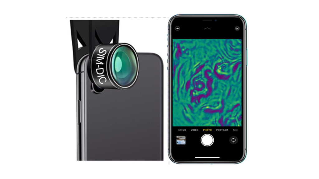
Hu’s plans to adapt a miniaturized DIC microscope to a smartphone. Credit: Guangwei Hu.
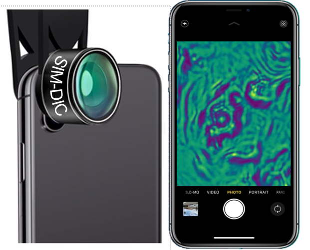
Hu’s plans to adapt a miniaturized DIC microscope to a smartphone. Credit: Guangwei Hu.
We use flat, ultrathin optical components called metasurfaces, which are sub-wavelength patterned layers that interact strongly with light. A single-layer metasurface composed of thousands of nanostructures with a few hundred nanometer thickness will generate two images with one slightly larger than the other, instead of the directional lateral shift of conventional DIC microscopes. Those two waves will interfere with each other, creating the image contrast at the focal point where the CCD is located.
Our recent work has demonstrated the feasibility of a single-shot, isotropic, and miniaturized DIC system. We have successfully imaged moving micro- and nano-particles to track their 3D movements, as well as unstained breast cancer tissues and cells. The next step is to see whether our device can serve as an add-on module for existing consumer electronics, such as smartphones or medical scopes.
A group effort
Other research groups are actively pursuing similar goals to ours, driven by rapid innovations in flat optics and metamaterials.
Federico Capasso at Harvard University is a renowned pioneer of using metasurfaces as a platform for flat optics. He has been actively working on a project with Melissa Suter at Mass General Research Institute to develop an endoscopic optical coherence tomography catheter for clinical applications, including detecting early-stage tumors in the lung. The nano-optic endoscope, which incorporates a flat metalens, boasts superior resolution and higher imaging depth of focus than traditional endoscopic optical imaging systems.
The lab of Andrei Faraon at Caltech has created a compact folded metasurface spectrometer that leverages a folded optics architecture with the help of metasurfaces. Optical spectrometry is a key technique in many fields, including environmental monitoring, materials analysis, medical diagnosis, and biological sensing. The approach significantly reduces the instrument’s size while also increasing the potential for integration into consumer electronics.
Mark Brongersma at Stanford University recently proposed a conceptually new approach to phase contrast imaging that harnesses the optical response of a specialized metasurface. Similar to DIC microscopy, phase contrast imaging allows for the visualization of translucent objects that are challenging to see in an ordinary light microscope.
Towards a brighter and transparent future
“Light-based technologies have a critical role to play in helping to solve the world’s problems, in diverse areas like sensing, communications, imaging, and computing.”
“Light-based technologies have a critical role to play in helping to solve the world’s problems, in diverse areas like sensing, communications, imaging, and computing.”
In the future, I anticipate that flat optical elements and metasurfaces will facilitate the development of smaller, more efficient, and more powerful microscopes. Ideally, we will create systems that match the capabilities of microscopes currently found only in laboratory settings, but condensed into a compact, portable, cost-efficient and add-on module form. Such devices can then be put in the hands of communities who need them most, to ensure their people are breathing clean air, drinking safe water, and staying healthy.
Light-based technologies have a critical role to play in helping to solve the world’s problems, in diverse areas like sensing, communications, imaging, and computing. I anticipate that their importance will only grow over time, meaning we need more people with different backgrounds to join in these efforts. The more people we have collaborating on optical solutions, the brighter and the clearer all of our futures will be.

Guangwei Hu is an early-career researcher at Nanyang Technological University, Singapore.
Explore other International Day of Light content from Optica

Read Optica President-Elect Jim Kafka's article "Lasers for Sustainability" also released in celebration of the 2024 International Day of Light.
