FIO plenary highlights: opportunities in optical imaging by wavefront shaping with spatial light modulators
FIO plenary highlights: opportunities in optical imaging by wavefront shaping with spatial light modulators
Ashley Collier, Corporate Communications Manager, Optica
Monika Ritsch-Marte, Medical University of Innsbruck
In her current role at the Medical University of Innsbruck, Ritsch-Marte oversees applied optics with a focus on laser applications & biomedical research. In her plenary presentation at Frontiers in Optics + Laser Science (FIO LS), Ritsch-Marte discussed opportunities in optical imaging by wavefront-shaping with Spatial Light Modulators (SLMs) and her recent work in Innsbruck.
She described the three types of devices of spatial light modulators: deformable mirrors, digital micromirror devices, and liquid crystal SLM (LC-SLM) as the three different tools for wavefront-shaping in optical imaging. She discussed the general advantages and disadvantages of the three device types.
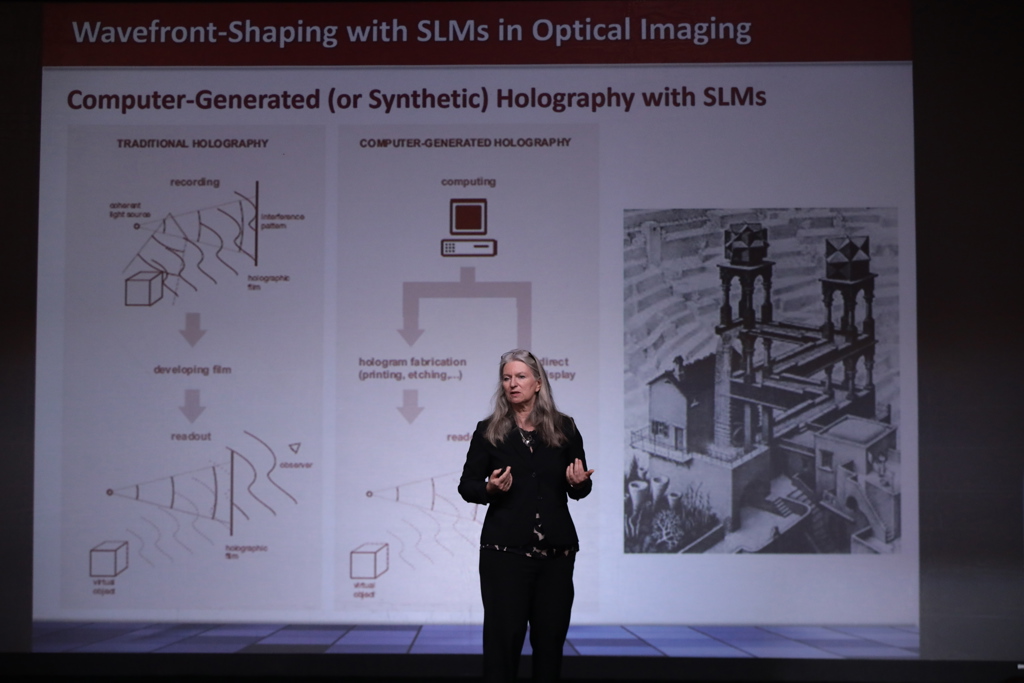
At the Medical University of Innsbruck, Ritsch-Marte described the Innsbruck group’s experience of working with liquid crystals displays for applications in holographic optical tweezers, beam shaping, advanced microscopy, optical imaging, aberration correction, and multiplexing into sub-images. She explained experiments performed in her lab, such as programmable-Fourier filtering with an SLM, which allows for phase contrast optimization and sample customization. Ritsch-Marte shared the advantages of computer-generated holography with SLMs compared to traditional holography.

Ritsch-Marte explained the strengths and limitations of LC-SLMs in terms of phase stroke, update speed, display resolution and diffraction efficiency, by means of examples drawn from her group’s 20-year experience. These covered SLM-controlled spiral phase-contrast, single-shot quantitative DIC, depth of field multiplexing, parallelized confocal imaging, and optoacoustic holography. She shared insights on the limitations caused by phase-only modulation and how one can nevertheless shape a natural wavefront by a ‘double-SLM technique’. She added the trade-off between field of view and resolution when multiplexing. She explained how color dispersion of LC-SLMs is often seen as a negative by-effect, but how this can be turned around and utilized for multi-colour synthetic holography.
Later in her presentation, Ritsch-Marte described the challenges of wavefront shaping for diffuse tissues. When correcting wavefront distortions, Ritsch-Marte compared the advantages and disadvantages of indirect (sensorless) and direct wavefront sensing. She shared the work done in Innsbruck with Alexander Jesacher to create DASH – Direct Adaptive Scattering Compensation Holography. She described DASH as a fast converging algorithm for scattering compensation in nonlinear microscopy. She also noted DASH required half as many photons to achieve a 50% correction and required less time to give a "very quick reasonable correction." Ritsch-Marte indicated how the DASH image improved after only one iteration in measurements on fluorescent beads behind a scattering layer. She also showed results of wavefront correction 500 µm deep into mouse brain tissue, clearly showing GFP-expressing microglia cells.
Moving forward, Ritsch-Marte looks ahead to continuing her exploration of biomedical research as she and her team will collaborate with the Physiology Institute at the Medical University of Innsbruck to study biological questions on the physiology of pain.
To view Monika Ritsch-Marte’s plenary presentations, visit the FIO LS website.
Possible References:
Maurer, C., Jesacher, A., Bernet, S., & Ritsch‐Marte, M. (2011). What spatial light modulators can do for optical microscopy. Laser & Photonics Reviews, 5(1), 81-101.
Jesacher, A., & Ritsch-Marte, M. (2016). Synthetic holography in microscopy: opportunities arising from advanced wavefront shaping. Contemporary Physics, 57(1), 46-59.
May, M. A., Barré, N., Kummer, K. K., Kress, M., Ritsch-Marte, M., & Jesacher, A. (2021). Fast holographic scattering compensation for deep tissue biological imaging. Nature Communications, 12(1), 1-8.
