Researchers integrate fast OCT system into neurosurgical microscope
About Optica
17 September 2024
Researchers integrate fast OCT system into neurosurgical microscope
Clinical study of microscope-integrated system lays groundwork for using OCT to define tumor margins and reveal subsurface brain anatomy
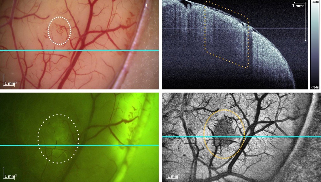
Caption: Researchers have integrated their MHz-OCT system into a neurosurgical microscope. The top left picture shows the typical surgical microscope view of a tumor below the brain’s surface illuminated with white light. With a fluorescent agent and viewed under blue-violet light, parts of the tumor become slightly visible (lower left). The new system can look below the surface to reveal the structure in depth as seen in the upper left image, which shows the slice indicated by the blue line. When projected into a top view as seen in the lower left picture, the tumor becomes clearly visible.
Credit: Universität zu Lübeck, Medizinisches Laserzentrum Lübeck GmbH & Universitätsklinikum Schleswig-Holstein
WASHINGTON — Researchers have successfully integrated a megahertz-speed optical coherence tomography (MHz-OCT) system into a commercially available neurosurgical microscope and demonstrated its clinical usefulness. This advancement represents an important step toward developing an OCT instrument that could be used to identify tumor margins during brain surgery.
“The MHz-OCT system we developed is very fast, about 20 times faster than most other OCT systems,” said Wolfgang Draxinger from Universität zu Lübeck. “This allows it to create 3D images that reach below the brain’s surface. These could be processed, for example with AI, to find and show parts that are not healthy and further need treatment yet would remain hidden with other imaging methods.”
In the Optica Publishing Group journal Biomedical Optics Express, researchers led by Robert Huber describe results from a clinical study of the microscope integrated MHz-OCT system. They showed that the system can be used during a surgical procedure to acquire high quality volumetric OCT cross-sectional scans within seconds, with the images immediately available for post-processing.
“We see our microscope integrated MHz-OCT system being used not just in brain tumor surgeries, but as a tool in every neurosurgery setting, since it can acquire high contrast pictures of anatomy such as blood vessels through the thick membrane that surrounds the brain,” said Draxinger, first author of the new paper. “This could significantly improve outcomes for procedures requiring detailed information about anatomical structures beneath the brain’s surface, such as deep brain stimulation for Parkinson’s disease.”
Speeding up OCT
The researchers have been working for some time to speed up OCT technology by improving the light sources and sensors used and developing software to process the large amount of data generated. This resulted in the development of an MHz-OCT system that can achieve over a million depth scans per second.
Caption: This video shows the first application of the microscope integrated MHz-OCT system in a clinical setting. The surgeon can be seen operating the microscope, and the scanning process is visible on the screens in the background.
Credit: Credit: Universität zu Lübeck, Medizinisches Laserzentrum Lübeck GmbH & Universitätsklinikum Schleswig-Holstein
The megahertz speed makes it possible to acquire over a million depth scans in just one second. This imaging speed is possible because the system incorporates a Fourier domain mode locking laser, first conceived in 2005 by Huber during his doctoral thesis research at MIT under James G. Fujimoto, who together with Eric Swanson and David Huang invented OCT. In addition, the development of graphics processing unit (GPU) technology over the past 15 years led to the computational capabilities required for processing the raw OCT signal into readable visuals without a bulky computer.
To find out if the MHz-OCT instrument they developed could be used to view brain tumor margins, the researchers integrated it with a special type of microscope that surgeons use to get a better view of the brain.
Taking it to the operating room
After building the integrated system, they tested it with calibration targets and tissue analog phantoms. Once satisfied with these results, they proceeded through patient safety testing and then began a clinical study investigating its application in brain tumor resection neurosurgery in 30 patients.
“We found that our system integrates well with the regular workflow in the operating room, with no major technological issues,” said Draxinger. “The quality of the images acquired surpassed our expectations, which were set low due to the system being a retrofit.”
During the clinical study, the researchers acquired about 10 TB of OCT imaging data along with matched pathological histology information. They note that they are still in the very early stages of understanding the data and images the new system is producing and developing AI methods for classifying the tissue. Thus, it will likely be years before this technology could be widely used to support brain tumor resection neurosurgery.
They are also preparing a study in which the new system will be used to demonstrate the exact location of brain activity in reaction to, for example, an external stimulus, during neurosurgery. This could hold promise for enhancing the precision of implantation of neuroprosthetic electrodes, allowing more accurate control of prosthetic devices by tapping into the brain's electrical signals.
Paper: W. Draxinger, N. Detrez, P. Strenge, V. Danicke, D. Theisen-Kunde, L. Schützeck, S. Spahr-Hess, P. Kuppler, J. Kren, W. Wieser, M.M. Bonsanto, R. Brinkmann, R. Huber, “Microscope Integrated MHz Optical Coherence Tomography System for Neuro-Surgery: Development and Clinical In-Vivo Imaging,” Biomed. Opt. Express, 15, 10, (5960), 2024 https://doi.org/10.1364/BOE.530976
About Optica Publishing Group
Optica Publishing Group is a division of the society, Optica, Advancing Optics and Photonics Worldwide. It publishes the largest collection of peer-reviewed and most-cited content in optics and photonics, including 18 prestigious journals, the society’s flagship member magazine, and papers and videos from more than 835 conferences. With over 400,000 journal articles, conference papers and videos to search, discover and access, our publications portfolio represents the full range of research in the field from around the globe.
About Biomedical Optics Express
Biomedical Optics Express serves the biomedical optics community with rapid, open-access, peer-reviewed papers related to optics, photonics and imaging in biomedicine. The journal scope encompasses fundamental research, technology development, biomedical studies and clinical applications. It is published monthly by Optica Publishing Group and edited by Christoph Hitzenberger, Medical University of Vienna, Austria. For more information, visit Biomedical Optics Express.
Media Contact
