Researchers detect hidden threats with advanced x-ray imaging
About Optica
23 May 2024
Researchers detect hidden threats with advanced x-ray imaging
Multi-contrast x-ray imaging combined with machine learning effectively distinguishes threatening materials in thousands of complex scenarios
WASHINGTON — Researchers have combined various x-ray imaging technologies to create multi-contrast images that can be used to detect threatening materials such as explosives in thousands of complicated scenarios. The new approach, which also leverages readily available machine learning procedures for materials classification, could be useful for security screening as well as applications in the life and physical sciences.

Credit: Thomas Partridge, University College London
“This method is particularly well suited to discriminating objects with very similar elemental composition,” said research team leader Thomas Partridge from University College London in the UK. “It could be used in airport security or any inline scanning operation to examine materials flagged as suspicious by an initial fast scan such as a traditional x-ray system.”
In Optica, Optica Publishing Group’s journal for high-impact research, the researchers show that the new approach was highly effective in accurately detecting and identifying explosives in almost 4,000 scans of threatening and non-threatening materials hidden inside bags or obscured by various types of objects. They achieved a near-perfect recall rate of 99.68%, with just one false negative, from the threat-bearing cases.
“Although more work is needed, this approach could also prove useful for medical imaging,” said Partridge. “While traditional x-ray imaging struggles to separate healthy from diseased tissue, other studies have suggested that phase contrast imaging might be able to capture textures that could be used to distinguish healthy and benign tissues.”
Unlocking material secrets
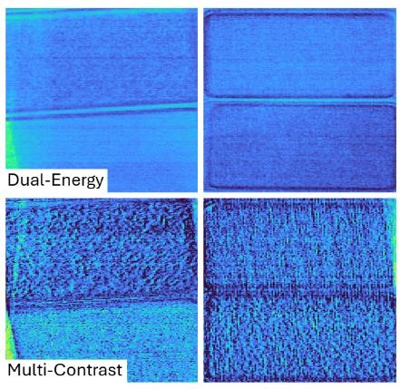
The x-ray machines found in airports or medical facilities are based on x-ray attenuation, which images the reduction in intensity in x-rays after they pass through a material. The new technique creates multi-contrast images by combining conventional x-ray attenuation data at various x-ray energies with x-ray phase information, which consists of refraction and dark-field channels.

Caption: The new technique produces multi-contrast images by combining conventional x-ray attenuation images at various x-ray energies (two energies shown on the top) with x-ray phase information, which consists of refraction and dark-field channels. This offers significantly better enhancement of textures and grains, as shown in the example composite image on the bottom, allowing the discrimination of materials with very similar elemental compositions.
Credit: Thomas Partridge, University College London
“Many explosives and common everyday items are composed of primarily carbon, hydrogen, nitrogen and oxygen, a similarity that makes them difficult to separate with x-ray attenuation alone,” said Partridge. “The additional channels offer significantly better enhancement of edges as well as textures and grains of materials, allowing the discrimination of objects with very similar elemental compositions.”
This work builds on previous efforts by the researchers to use multi-contrast x-ray phase enhanced imaging with machine learning approaches for threat detection with a smaller number of explosives and benign objects.
In the new experiment, they significantly scaled up the number of materials investigated and the number of imaging scenarios to better mimic real world situations. They also created a more effective scanning system with a resolution that could be changed by modifying the scanning speed and applied edge illumination phase contrast. Edge illumination involves placing masks before and after the sample to create the sub-pixel x-ray ‘beamlets’ needed to make the system sensitive to phase signals. One key advantage of this illumination approach is that it works with incoherent x-ray sources, widening its applicability.
Since the increased complexity of the imaging scenarios required more sophisticated protocols, the researchers applied machine learning with a hierarchical architecture that separated the cluttering objects before distinguishing material types. This made it possible to rapidly discern subtle differences in shapes and textures to distinguish materials based on key identifying features.

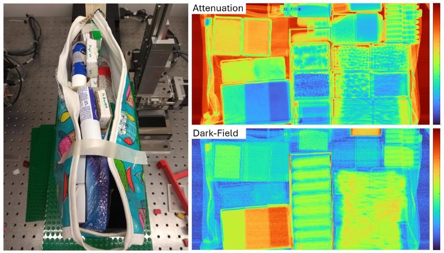
Caption: The researchers tested the new technique with 19 threat materials and 56 non-threat materials, some of which are pictured.
Credit: Thomas Partridge, University College London
Detecting threats
To test the new technique, they used 19 threat materials and 56 non-threat materials, all at three thicknesses and obscured by a range of cluttering objects such as brushes, facewipes, socks and other items passengers would have in a carry-on bag. By using all the acquired contrast channels, the researchers demonstrated not just material discrimination but identification in some cases. Using deep learning to analyze the signals from the combination of x-ray contrasts provided very promising results, with just a single miss out of 313 threat cases.
The researchers say that translating this approach to a commercial setting would require improving the scanning speed through further optimization of the system. The robustness of material discrimination also needs to be tested on a larger set of data. One area of active study for the team is combining the method with 3D computed tomography scanning, which is being explored for security purposes because of its ability to provide detailed, three-dimensional images of objects.
Paper: T. Patridge, S.S. Shankar, I. Buchanan, P. Modregger, A. Astolfo, D. Bate, A. Olivo, “Multi-contrast x-ray identification of inhomogeneous materials and their discrimination through deep learning approaches,” 11, 6, 759-767 (2024).
DOI: doi.org/10.1364/OPTICA.507049
About Optica Publishing Group
Optica Publishing Group is a division of the society, Optica, Advancing Optics and Photonics Worldwide. It publishes the largest collection of peer-reviewed and most-cited content in optics and photonics, including 18 prestigious journals, the society’s flagship member magazine, and papers and videos from more than 835 conferences. With over 400,000 journal articles, conference papers and videos to search, discover and access, our publications portfolio represents the full range of research in the field from around the globe.
About Optica
Optica is an open-access journal dedicated to the rapid dissemination of high-impact peer-reviewed research across the entire spectrum of optics and photonics. Published monthly by Optica Publishing Group, the Journal provides a forum for pioneering research to be swiftly accessed by the international community, whether that research is theoretical or experimental, fundamental or applied. Optica maintains a distinguished editorial board of more than 60 associate editors from around the world and is overseen by Editor-in-Chief Prem Kumar, Northwestern University, USA. For more information, visit Optica.
Aaron Cohen
(301) 633-6773
aaroncohenpr@gmail.com
